
Eyelids protect your eyes from any foreign bodies while keeping them lubricated throughout. Any alteration in the shape, position or function of your eyelids can predispose your eyes to a plethora of ailments or interfere with our vision.
Our eyelid is a complex structure consisting of three theoretical layers:
Eyelid malpositions include any unnatural or incorrect positioning and orientation of eyelids due to various factors that influence any of the three layers of the eyelids. They could be caused due to ageing, trauma, scarring, birth defects or medical disease involving any or all of the three layers.
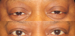
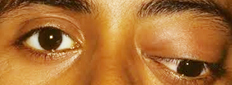
Ptosis – Ptosis is drooping of the upper lid. It may be present by birth, or appear later in life. Ptosis occurs when the muscles that raise the eyelid (levator and Müller's muscles) are not strong enough to do so perfectly. It can affect one eye or both eyes and occurs more often in the elderly, as muscles in the eyelids might possibly begin to deteriorate.
Ptosis can be caused by the weakness of aponeurosis of the levator muscle, nerve abnormalities, trauma, inflammation or lesions of the lid or orbit.
Surgical procedures include:
Non-surgical modalities like the use of "crutch" glasses or special contact lenses to support the eyelid might possibly also be used
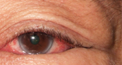
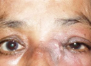
It is a medical term used to describe sagging and outward turning of the lower eyelid and eyelashes. This rubbing can lead to excessive tearing, crusting of the eyelid, mucous discharge and irritation of the eye
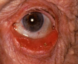
It is a medical term used to describe sagging and outward turning of the lower eyelid and eyelashes. . This rubbing can lead to excessive tearing, crusting of the eyelid, mucous discharge and irritation of the eye.
Evaluation and treatment of eyelid malpositions needs to be disease-specific and tailored to the patient’s needs. This requires consultation with an eye surgeon familiar with treating various eyelid malpositions.
Trichiasis: Lashes pointing to the eye
Cancer of the eyelid, like any other cancer, can be a worrying thing. Treatments are variable and depend on the type of cancer. Broadly classified, eyelid tumors can be of the following types –
These include epidermoid cysts, dermoid cysts, sweat ductal cysts and epidermal inclusion cysts.
As they are benign, they are easily treatable, though the treatment option for each varies.
While technically these are not tumors, they must be borne in mind when making a diagnosis of cancer. Lesions include hordoleum (stye), chalazion and parasitic infections.
Stye often need antibiotics and surgical drainage, chalazion benefits from simple observation and hot compresses if required, along with a gentle massage, while parasitic infections could need anti-parasitic medication.
Hemangiomas are vascular lesions that can mimic eye tumors. They could be a capillary hemangioma, cavernous hemangioma or a lymphangioma.
They are often seen in infants and children. If found, it needs fairly urgent treatment as it can have an impact on vision and result in blindness.
Treatments include steroids (administered as a ointment or injection), laser photocoagulation and surgical excision if required.
These include squamous papillomas, seborrhoeic keratosis, inverted follicular keratosis and keratoacanthoma. Squamous papillomas appear round or pedunculated and have a smooth surface.
They can be removed surgically or in some cases interferon could be used. Seborrhoeic keratosis is often monitored for a change in shape or size before any treatment is considered. They often need a biopsy to confirm their benign nature.
A number of different eyelid lesions could become cancerous. They include Actinic keratosis, leukoplakia, Xeroderma pigmentosum and radiation dermatosis.
Actinic keratosis occurs in sun-exposed areas and appears like a white, scaly lesion. Excision biopsy aids diagnosis.
Common viral lesions include molluscum contagiosum, verruca vulgaris , herpes simplex and herpes zoster.
Molluscum contagiosum is seen in individuals with low immunity, and can cause conjunctivitis. They are treated with cryotherapy or excision. Herpes simplex and zoster are treated with antiviral agents.
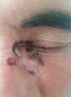
These are the cancerous lesions, and include basal cell carcinoma, squamous cell carcinoma, malignant melanoma and sebaceous gland carcinoma.
Basal cell carcinoma is the most common eyelid malignancy, and occurs as a small module at the inner aspect of the eye (medial canthus). They are locally invasive and require surgical excision.
Squamous cell carcinoma is not as common, and appears red and scaly with an ulcer in the center. It is prone to distant spread so must be excised as soon as possible.
Sebaceous carcinoma is seen in elderly population, and can spread to lymph nodes and distant organs. Radical surgery is often needed. Melanomas are rare and could need excision.
These include Xanthelasma, nevi and Caruncular Tumors. Most are benign and require simple treatments. Xanthalesma is associated with high cholesterol levels in the blood. Nevi need excision if they change size or shape.
Closed globe injury or Non-penetrating trauma: The eye globe is intact, but the seven rings of the eye have been classically described as affected by blunt trauma.
Penetrating trauma: The globe integrity is disrupted by a full-thickness entry wound and may possibly be associated with prolapse of the internal contents of the eye.
Perforating trauma: The globe integrity is disrupted in two places due to an entrance and exit wound (through and through injury). This is a quite severe type of eye injury.
Blowout fracture of the orbit is caused by blunt trauma, classically described for fist or ball injury, leading to fracture of the floor or medial wall of the orbit due to sudden increased pressure on the orbital contents. A fracture in the bones around the eye can cause double vision, and a sunken small appearance of the eye. The fracture is repaired with an implant or plate. The best results are obtained in surgery within 2 weeks of injury, but surgery can also be done later.
Quick attention to the injury as well as possible need for plastic and reconstructive surgery are important to restore the proper function as well as cosmetic appearance.
If the eye is damaged to the extent that it must be removed, an artificial eye may possibly inserted. There is no way to restore vision to the eye in this instance.
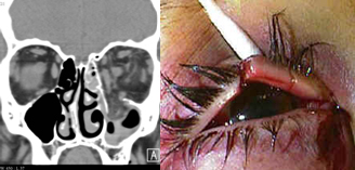
It is any tumor that occurs within the orbit of the eye. The orbit is a bony housing in the skull about 2 inches deep that provides protection to the entire eyeball except the front surface. It is lined by the orbital bones and contains the eyeball, its muscles, blood supply, nerve supply, and fat.
Tumors might possibly develop in any of the tissues surrounding the eyeball and might possibly also invade the orbit from the sinuses, brain, or nasal cavity, or it might possibly metastasize (spread) from other areas of the body. Orbital tumors can affect adults and children. Fortunately, most are benign.
Most childhood orbital tumors are benign and are the result of developmental abnormalities.Common orbital tumors in children are dermoids (cysts of the lining of the bone) and hemangiomas (blood vessel tumors).Malignant tumors are unusual in children, but any rapidly growing mass should be cause for concern.Rhabdomyosarcoma is the most common malignant tumor affecting children, and it usually occurs between the ages of 7 and 8.
The most common orbital tumors in adults are also blood vessel tumors, including hemangioma, lumphangioma, and arteriovenous malformation.Tumors of the nerves, fat, and surrounding sinuses occur less often.Lymphomas are the most commonly occurring malignant orbital tumors in adults.
Metastic tumors most commonly arise from the breast and prostate, while squamous and basal cell cancer can invade the orbit from surrounding skin and sinus cavities.
Symptoms of an orbital tumor might possibly include
Prominence of the eyes is not necessarily the result of a tumor, but might possibly result from inflammation such as that caused by Graves' thyroid disease.
In children, parents might possibly first notice a droopy eyelid or slight protrusion of the eye.
Orbital tumors are most commonly diagnosed with either a CAT scan or MRI. If either of those tests look suspicious, a biopsy might possibly be performed.
Treatment of orbital tumors varies depending on the size, location, and type.Some orbital tumors require no treatment, while others are best treated medically or with the use of radiation therapy.
Some might possibly need to be totally removed by either an orbital surgeon or a neurosurgeon, depending on the particular case. After removal, additional radiation or chemotherapy might possibly be required.
Surgery has become much safer because CT scans and MRI testing can help pinpoint the location and size of the tumor.
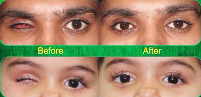
Enucleation/ evisceration– A highly damaged eye may be replaced with an orbital implant; this restores the comfort and appearance.
Socket reconstruction– where the eye has been removed for a serious disease, the socket of the eye may get shrunken. The socket is reconstructed to improve comfort and appearance.
Ocularistry: When an eye has been removed due to disease, it may have a serious impact on the psychological and social well-being of the patient. Ocularistry is the art and science of manufacturing an artificial eye. For customised prosthesis (tailor-made artificial eye) measurements are taken from the patient’s own eye, and colour match is done. Thus the prosthesis fits well, is very comfortable, and looks similar to the patient’s own eye.
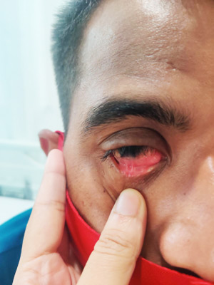
Symptoms: acute onset, tearing (from nasolacrimal duct obstruction), redness, purulent discharge, tender swollen lacrimal sac. An infection within the "tear duct" causes a painful swelling in the inner corner of the eyelids.
If the tearing causes severe symptoms, surgery can be performed to create a new tear duct. This operation is called "dacryocystorhinostomy. Small silicone tubes may be placed in the tear system to keep the new tear duct open while healing occurs.
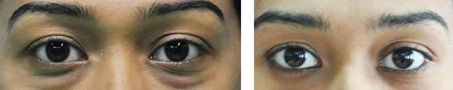
Your eyes including your eyelids, are perhaps one of the first things people notice in you. This makes your eyes and eyelids one of the most important components for an appealing facial expression and aesthetic appearance. Any visible change in the shape or size of the orbital or periorbital region can spoil the look of your face.
As you age and grow older, your eyelids may possibly become ‘droopy’ or ‘baggy’ due to the stretching of your eyelid skin and gradually decreasing tone of your eyelid muscles. Your droopy eyelids and brow together cut a sorry figure for your face making you look tired, sleepy and haggard, further leading to eyelid or brow straining or both. In extreme cases, your saggy, baggy eyelids can even obstruct your vision, particularly peripheral vision causing difficulty in reading or driving.
Blepharoplasty ensures cosmetic or functional corrections to the area around your eyes to improve your look or to correct any abnormalities in function.
Blepharoplasty involves removal or repositioning of excess tissue as well as reinforcement of surrounding muscles and tendons to reshape the upper eyelid, lower eyelid or both.
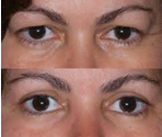
It assists in the reduction of excess skin and fat in the upper eyelids.
Thyroid eye disease: Thyroid disease can have a serious effect on the eye. The eye develops a bulging look; the patient can have itching and burning, discomfort, corneal ulcers, double vision, and even loss of vision.
Our thyroid eye disease clinic monitors the eye health regularly, so that any danger to the eye is prevented. By injection and surgery, the changes done by the disease can be reversed back to normal.
Botox clinic hemifacial spasm, essential blepharospasm, wrinkle treatment. Due to over-action of muscles around the eye, the eyes can go into spasm – where the face and eyes twitch; patient is not able to open the eyes comfortably. This can be treated safely and effectively by injection of botulinum toxin.
Botulinum toxin can also be used to treat wrinkling and aging around the eye.
Lorem Ipsum is simply dummy text of the printing and typesetting industry. Lorem Ipsum has been the industry's standard dummy text ever since the 1500s
Lorem Ipsum is simply dummy text of the printing and typesetting industry. Lorem Ipsum has been the industry's standard dummy text ever since the 1500s
Lorem Ipsum is simply dummy text of the printing and typesetting industry. Lorem Ipsum has been the industry's standard dummy text ever since the 1500s
Near Olympus, CH Baktawar Singh Rd, Sector 38, Gurugram, Haryana 122001
Copyright © 2022 Dr. Svati Bansal All Rights Reserved.
Healthcare Web Design Agency Medkeon